RGUHS Nat. J. Pub. Heal. Sci Vol: 15 Issue: 4 eISSN: pISSN
Dear Authors,
We invite you to watch this comprehensive video guide on the process of submitting your article online. This video will provide you with step-by-step instructions to ensure a smooth and successful submission.
Thank you for your attention and cooperation.
1Dr. Aditya J Baindur, Department of Pediatric Surgery, Sawai Man Singh Medical College & SPINPH, Jaipur, Rajasthan, India.
2Department of Pediatric Surgery, Sawai Man Singh Medical College & SPINPH, Jaipur, Rajasthan, India
3Department of Pediatric Surgery, Sawai Man Singh Medical College & SPINPH, Jaipur, Rajasthan, India
4Department of Pediatric Surgery, Sawai Man Singh Medical College & SPINPH, Jaipur, Rajasthan, India
*Corresponding Author:
Dr. Aditya J Baindur, Department of Pediatric Surgery, Sawai Man Singh Medical College & SPINPH, Jaipur, Rajasthan, India., Email: adityajbaindur@gmail.com
Abstract
Fetus in fetu is a rare congenital anomaly, with an incidence of approximately 1 in 5,00,000 live births. A 5-month-old pediatric patient presented with gross abdominal distension and respiratory distress. Contrast-enhanced CT of the abdomen revealed a well-encapsulated, multiloculated solid-cystic lesion in the right retroperitoneum, containing intralesional fatty components and calcifications forming appendiceal bones. Exploratory laparotomy was performed, and the lesion was excised in toto. The patient remains under regular follow-up. Given the rarity of fetus in fetu, we present this case to contribute to the literature and provide further insights into this unusual condition.
Keywords
Downloads
-
1FullTextPDF
Article
Introduction
Fetus in fetu (FIF) is a rare congenital anomaly. The incidence of FIF roughly corresponds to 1 in 5,00,000 live births.1 Historically, it can be traced back to the German Anatomist - Johann Meckel for describing the condition as early as 18th century.2,3 It is comprised of a malformed twin incorporated into the surviving twin, usually presenting as an abdominal mass. Unequal division of totipotent cells in the blastocyst of diamniotic monozygotic twins leads to the formation of a small cluster of cells that develop into a mass. Subsequent inclusion of this cell cluster into the co-twin results in the formation of a vestigial, teratomatous remnant. The majority of reported cases (80%) are located in the retroperitoneum.4 However, rare occurrences have also been documented in unusual sites like the cranium and scrotum.5 As fetus in fetu is itself an exceedingly rare entity, with only around 200 cases reported to date.6 With this background, we present this case to contribute to the existing literature and provide further insight into this unusual condition.
Case Report
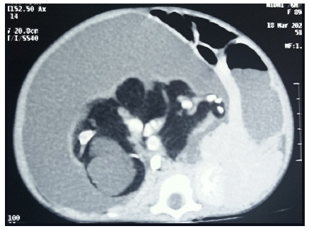
A 5-month-old female infant presented with gross abdominal distension associated with respiratory distress. The parents reported the presence of an abdominal mass since birth, which had gradually progressed. The child was born at full term via caesarean section without any complications. There was no history of twin birth in the family. On examination, the child exhibited hurried breathing with distress and marked abdominal distension. A firm, non-tender, well-defined spherical mass was palpable, occupying the upper and lower quadrants of the right side of the abdomen and extending across the midline. Routine blood investigations were within normal limits. Serum alpha-feto protein (AFP) levels were elevated, while serum beta-human chorionic gonadotropin (HCG), carcinoembryonic antibody (CEA) levels were within normal range. Abdominal ultrasonography revealed a large cystic mass with internal solid components resembling limb buds in the right retroperitoneal region. The bowel loops were found to be pushed anteriorly. Contrast-enhanced computed tomography (CECT) of the abdomen revealed a well-encapsulated, multi-loculated solid-cystic lesion containing intralesional fat components and calcifications forming appendiceal bones in the right retroperitoneum (Figure 1). Based on the presence of limb buds and appendiceal bones, fetus in fetu was considered the most likely diagnosis, with a highly organized teratoma kept as a differential.
Treatment
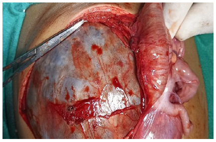
An exploratory laparotomy was performed, revealing a cystic mass extending superiorly from the inferior surface of the liver to the pelvic brim inferiorly. Laterally, the mass abutted the lateral abdominal wall, while medially it crossed the midline. It originated from the retroperitoneum, pushing the bowel loops anteromedially and right kidney anteroinferiorly (Figure 2). Posteriorly, it was abutting the portal vein and aorta. The mass received its blood supply from branches of the lower abdominal aorta. The lesion was excised in toto after careful dissection and ligation of the feeding vessels.
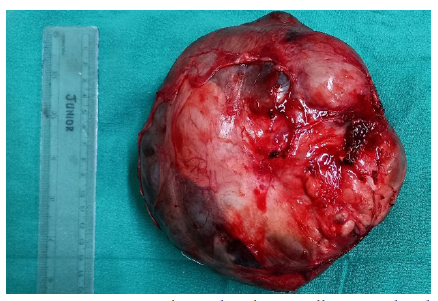
On gross examination, the excised specimen was a well-encapsulated mass with solid and cystic components, measuring approximately 16 x 18 x 14 cm (Figure 3). Upon opening the capsule, dirty brownish fluid was noted within the cystic component. The solid component comprised of malformed limbs with vestigial fingers, hair, skin, and orbit-like-projections. Cyst lining showed connection with the fetus through a tubular-like structure, resembling intestine with oedematous mucosal tissue folds (Figure 4). On sectioning the dorsal glistening surface, a well-developed vertebral column was also identified.
Discussion
Fetus in fetu (FIF) is a rare congenital anomaly with a malformed twin incorporated into the surviving twin. It is rarer compared to teratomas; hence, the possibility of teratoma cannot be ruled out initially. Thus, the knowledge of the Willis criteria is of utmost help. Willis emphasized that this entity should be defined as a mass containing a vertebral axis along with other formed organs or limbs.7 Our case meets these criteria; hence the diagnosis. However, there are authors who are in favour of considering this entity as a highly organized teratoma.8
Two theories address the development of fetus in fetu (FIF). One is the parasitic twin theory, which suggests that a malformed fetus gets incorporated into the body of its twin during the early intrauterine period and derives a common blood supply.9 The other theory proposes that the entity represents a highly organized teratoma.10 However, it is unusual to observe well-developed organs and vertebral bodies in a teratoma.11
Radiological examination plays a key role in the preoperative diagnosis. In our case, sonography and CT scan findings were helpful in the preoperative evaluation, raising a high suspicion of FIF. However, the intraoperative findings and examination of the specimen led to the confirmation of the diagnosis. Hence, we believe that exploratory laparotomy plays a key role in the management, as this rare pathology was confirmed only after surgical excision and specimen evaluation in our case.
Fetus in fetu is usually a benign condition. The symptoms are mainly due to mass per abdomen, which may compress bowel structures and lead to obstruction. In the present case, however, the child was brought in with respiratory distress, likely attributable to gross abdominal distension causing restricted diaphragmatic movement. There are some reports describing malignant features in fetus in fetu.1 Malignant transformation of residual tissue has also been documented.12 Hence, complete excision in toto is essential, and regular follow-up is required. Our patient remains under regular follow-up, subsequent scans show no signs of residual disease or recurrence.
Fetus in fetu is a rare entity, usually benign in nature. The presenting symptoms are primarily due to the space-occupying effect of the lesion. Imaging plays a key role, especially in identifying vertebral bodies, which can help confirm the diagnosis. Complete excision is essential, as it provides symptomatic relief and reduces the risk of future malignant transformation.
Conflict of Interest
Nil
Supporting File
References
1. Hopkins KL, Dickson PK, Ball TI, et al. Fetus-in-fetu with malignant recurrence. J Pediatr Surg 1997;32(10):1476-9.
2. Federici S, Prestipino M, Domenichelli V, et al. Fetus in fetu: Report of an additional, well-developed case. Pediatr Surg Int 2001;17:483-5.
3. Massad MG, Kong L, Benedetti E, et al. Dysphagia caused by a fetus-in-fetu in a 27-year-old man. Ann Thorac Surg 2001;71:1338-41.
4. Gupta SK, Singhal P, Arya N. Fetus-in-fetu: A rare congenital anomaly. J Surg Tech Case Rep 2010;2:77-80.
5. Sutthiwan P, Sutthiwan I, Tree-trakan T. Fetus in fetu. J Pediatr Surg 1983;18(3):290-2.
6. Arlikar JD, Mane SB, Dhende NP, et al. Fetus in fetu: two case reports and review of literature. Pediatr Surg Int 2009;25(3):289-92.
7. Willis RA. The structure of teratoma. J Pathol Bacteriol 1935;40:1-36.
8. Heifetz SA, Alrabeeah A, Brown BS, et al. Fetus in fetu: A fetiform teratoma. Pediatr Pathol 1988;8:215-26.
9. Chua JHY, Chui CH, Prasad TRS, et al. Fetus-in-fetu in the pelvis. Ann Acad Med Singap 2005;34:646-9.
10. Basu A, Jagdish S, Iyengar KR, Basu D. Fetus in fetu or differentiated teratomas? Indian J Pathol Microbiol 2006;49:563-5.
11. Kim OH, Shinn KS. Postnatal growth of fetus-in-fetu. Pediatr Radiol 1993;23(5):411-2.
12. Yaacob R, Zainal Mokhtar A, Abang Jamari DZH, et al. The entrapped twin: a case of fetus-in-fetu. BMJ Case Rep 2017;2017:bcr-2017-220801.


