RGUHS Nat. J. Pub. Heal. Sci Vol: 15 Issue: 4 eISSN: pISSN
Dear Authors,
We invite you to watch this comprehensive video guide on the process of submitting your article online. This video will provide you with step-by-step instructions to ensure a smooth and successful submission.
Thank you for your attention and cooperation.
1Dr. Ram Kumar Kaushik, Associate Professor, Department of Anatomy, Subharti Medical College, Swami Vivekanand Subharti University, Meerut, Uttar Pradesh, India.
2Department of Anatomy, Subharti Medical College, Swami Vivekanand Subharti University, Meerut, Uttar Pradesh, India
3Department of Anatomy, Subharti Medical College, Swami Vivekanand Subharti University, Meerut, Uttar Pradesh, India
4Department of Anatomy, Subharti Medical College, Swami Vivekanand Subharti University, Meerut, Uttar Pradesh, India
5Department of Anatomy, Subharti Medical College, Swami Vivekanand Subharti University, Meerut, Uttar Pradesh, India
6Department of Anatomy, VMMC and Safdarjung Hospital, Indraprastha University, New Delhi, India
*Corresponding Author:
Dr. Ram Kumar Kaushik, Associate Professor, Department of Anatomy, Subharti Medical College, Swami Vivekanand Subharti University, Meerut, Uttar Pradesh, India., Email: rkkaushik1258@gmail.com
Abstract
Background: The kidneys play a critical role in blood filtration, waste removal, and fluid and electrolyte balance, receiving approximately 20% of cardiac output through the renal arteries. These arteries divide into five segmental branches at the hilum: apical, upper, middle, inferior, and posterior.
Aim: The present study aimed to analyze the anatomical patterns of the upper segmental renal arteries using the corrosion cast technique.
Methods: This observational study was conducted in the Department of Anatomy between September 2019 and February 2024. Thirty pairs of kidneys were obtained from cadavers during routine dissections conducted by medical students. Renal artery corrosion casts were prepared by injecting cellulose acetate butyrate into the abdominal aorta, followed by solidification and maceration of the specimens. The observations were analyzed to identify the prevalence, origin, and variations of the upper segmental renal arteries. Statistical analysis was carried out using the Chi-square test.
Results: At the kidney hilum, the renal artery divides into anterior and posterior branches. In 10% of cases, the upper segmental artery originated directly from the renal artery. The anterior division was the most frequent source of the upper segmental artery (66.6%), with a posterior division origin in 3% of cases. In 15% of specimens, the upper segmental artery originated alongside the apical artery from the anterior division.
Conclusion: This morphological study of the origin, course, variations, and distribution of the upper seg-mental renal arteries provides valuable insights for renal transplantation, as well as for medical, surgical, and radiological applications.
Keywords
Downloads
-
1FullTextPDF
Article
Introduction
The current study aimed to examine variations in the upper segmental renal artery, which are crucial for medical professionals such as surgeons and radiologists. Recognizing these anatomical differences has significant implications for procedures including renal transplantation, abdominal aortic aneurysm repair, radical nephrectomy, and the management of renovascular hypertension. An In-depth understanding of these variations can improve surgical outcomes and facilitate the effective management of renal conditions, ultimately enhancing patient care.
Knowledge of variations in renal segmental artery anatomy is important during partial nephrectomy, as it facilitates a bloodless surgical approach and preserves renal segments. To achieve a bloodless field and reduce ischemic damage to the organ, renal branches can be selectively clamped according to the kidney’s segmental structure.1 Renal cell carcinoma is among the most common malignancies, and kidney transplantation has become increasingly important and frequent.2,3 Proper management of these arteries during surgery can significantly impact the success of both cancer treatments and transplant procedures.
The renal arteries develop from the lateral branches of the abdominal aorta during embryogenesis. Initially, multiple pairs of arteries supply the developing kidneys, but most regress, leaving one main renal artery on each side. Variations in the origin and branching patterns are common due to complex embryologic development. These variations can result in differences in the origin, number, and course of the upper segmental artery. Usually each kidney is vascularized by single renal artery originating from the abdominal aorta. At the hilum, this artery branches into anterior and posterior divisions, which is further subdivided into posterior, lower, middle, upper and apical segmental arteries. The upper segment receives its blood supply from upper segmental renal artery, a branch of the anterior division of renal artery.
During procedures such as calculus removal from the calyx, variations in arterial patterns can affect surgical outcomes. Grave's work on intrarenal arterial distribution established the foundation for understanding these patterns, dividing the renal parenchyma into five segments.4 Subsequent studies by Verma, Chatterjee, Ajmani, and Longia further explored these variations, reporting additional anatomical differences.5-8 Recognizing such variations is crucial in procedures including partial nephrectomy, renal artery embolization, kidney transplantation, and surgeries for renal trauma and renal artery stenosis, as it helps ensure successful outcomes and minimize complications.9 Using the corrosion cast method, the present study aimed to facilitate a bloodless surgical approach and preserve healthy renal segments during partial nephrectomy. Knowledge of upper segmental artery variations is essential to achieving these objectives.
Materials and Methods
This was an observational anatomical study, concentrating exclusively on the observation and detailing of anatomical features. The focus was on the upper segmental artery due to its unique path and crucial supply to about one-third to one-half of the kidney, providing essential insights for targeted surgical and clinical practices and laying a foundation for future studies on segmental renal arteries.
Ethical Considerations
Consent for the use of cadavers in this study was obtained through the institutional cadaver donation process. All cadaveric specimens used in this study complied with ethical guidelines and institutional protocols.
Sample Size
A total of 60 (thirty pairs) cadaveric human kidneys were used for the study.
Inclusion Criteria
1. Kidneys with associated segments of the abdominal aorta, ureter, inferior vena cava, and renal vessels.
2. Specimens appropriate for the corrosion casting technique.
Exclusion Criteria
1. Specimens with significant damage or deterioration affecting the integrity of the associated vessels.
2. Kidneys presenting with congenital malformations.
3. Specimens with previous surgical interventions.
4. Kidneys with signs of pathological conditions
Thirty pairs of cadaveric human kidneys were collected from the Department of Anatomy during routine anatomy dissections conducted by medical students. The study was carried out between September 2019 and February 2024.
Each kidney was removed along with the associated segments of the abdominal aorta, inferior vena cava, ureter, and renal vessels. The renal arteries in all specimens were examined for their gross anatomical features, and the relevant data were recorded for further statistical tests.
Stock solutions of red cellulose acetyl butyrate at a 20% concentration were prepared in advance by dissolving granules in acetone and used as needed. The solutions were stored in airtight glass jars for twelve hours to ensure uniform consistency. The kidneys were thoroughly rinsed under running tap water for approximately one hour followed by flusing through a cannula inserted into the upper part of the abdominal aorta after ligating the lower end. Warm normal saline was injected through the cannula to flush out blood clots.
An amount of red cellulose acetyl butyrate solution ranging between 25 and 50 cc was slowly injected into the upper end of the abdominal aorta using a 10 cc syringe and a wide bore cannula. The aorta was then securely ligated at both cut ends.
The infused specimens were submerged in covered trays containing 10% formalin for 72 hours. After fixation, the formalin was poured off, and the renal capsules were removed. The decapsulated specimens were then immersed in glass jars containing hydrochloric acid and left undisturbed for one week. This process macerated the renal tissue while preserving the solidified renal arteries.
Each individual cast obtained from the process was immersed in a 50% glycerin solution for approximately thirty minutes. Relevant observations were then made, focusing on the patterns and variations of the upper segmental, and other renal arteries. These findings were subsequently analyzed to identify specific trends and anomalies in the upper segmental renal arteries.
Results
The upper segmental artery was observed to be supplying the upper part of the central area on the anterior aspect of the kidney in all specimens. The high prevalence of this artery underscores the importance of recognizing its variations during surgical planning.
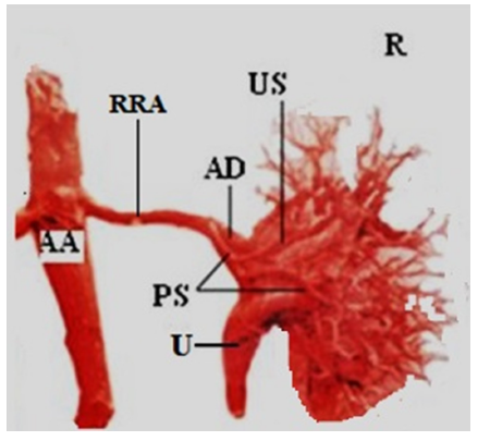
Five distinct branching patterns of the upper segmental artery were identified (Table 1). In 40 specimens (66.6%), the artery originated from the anterior division of the renal artery. It originated from the anterior division along with the apical artery in nine specimens (15%). In three specimens (5%), it arose from the middle segmental artery. The posterior segmental artery was the source of origin in two specimens (3%) (Figure 1). In six specimens (10%), the upper segmental artery arose from the main renal artery.
The Chi-square test was applied to analyze the distribution of the origins of the upper segmental renal artery, evaluating whether the observed frequencies differed significantly from the expected distribution. In this study, a Chi-square value of 84.16 with 4 degrees of freedom and a P-value of <0.05 indicated a highly significant difference, leading to the rejection of the null hypothesis.
Graves FT studied corrosion casts of kidneys and demonstrated that the renal arteries exhibit a segmental distribution intrarenally. Variations in the arterial pattern of the upper segmental artery have been reported by several authors. The origin of the upper segmental artery was found to be variable in studies by Fine H & Keen EN, Daescu E et al., Rani N et al., Mishra GP et al., and Lucas A Martin et al.10-14 A comparative summary is presented in Table 2.
Five different types of upper segmental renal arteries were observed depending on their origin, consistent with previous studies. Type I originated from the anterior division, type II from the anterior division along with the apical artery, type III from the anterior division with the middle segmental artery, type IV from the posterior segmental artery, and type V from the renal artery.
In the present study, the upper segmental artery most frequently (66.6%) originated from the anterior division of the renal artery (Type I), which is consistent with the findings reported by Fine H & Keen EN, Rani N et al., Mishra GP et al., and Lucas A Martin et al.10,12-14 In contrast, Type I was the second most common in the study by Daescu E et al., and was not observed in the study by Rani N et al.11-12
In the present study, the next most frequent type was Type II (15%), in which the upper segmental artery originates from the anterior division along with the apical artery. This finding is consistent with the results of Mishra GP et al., who reported a frequency of 15%.13
Type III, in which the artery arises from the anterior division with the middle segmental artery, was found only in 5% of specimens in the present study, aligning with the 1.67% reported by Daescu E et al.11 However, Mishra GP et al., reported a comparatively higher frequency of Type III (28%), while Rani N et al., did not observe this type.12-13
Type IV, originating from the posterior segmental artery, was reported at a relatively high frequency (10%) by Mishra GP et al.13 In the study by Daescu E et al., it was observed in 1.67% of cases, similar to the present study, which found a frequency of 3.3%.11 Type V, arising directly from the renal artery, was reported at a notably high frequency (40%) by Rani N et al.12 However, in the present study, it was less frequent (10%). This type was not observed in the studies by Fine H & Keen EN, Daescu E et al., Mishra GP et al., and Lucas A Martin et al.10,11,13,14
Discussion
In the present study, the upper segmental artery most frequently originated from the anterior division of the renal artery, aligning with previous anatomical studies, highlighting the typical vascular pattern observed in the renal artery system. The next most common origin involved the anterior division along with the apical artery, a variation crucial for surgeons and interventional radiologists during renal procedures to avoid complications and ensure effective outcomes. Notably, the upper segmental renal artery arising directly from the main renal artery was relatively less frequent, was relatively less frequent, underscoring the importance of recognizing anatomical variations.
The rare variation of the upper segmental artery originating from the posterior segmental artery carries significant surgical implications. Recognizing this variation can optimize the choice of surgical approach, reduce intraoperative risks, and enhance the precision of arterial clamping during nephron-sparing surgeries. Advanced preoperative imaging is essential for identifying such variations and tailoring surgical strategies accordingly. Understanding these variations aids in preoperative planning and intraoperative decision-making, providing a foundation for future studies on segmental renal arteries and improving clinical practices in renal surgeries and interventions.
While this study provides significant insights into the upper segmental renal artery, further research is needed to explore the full spectrum of anatomical variations and their clinical implications.
Conclusion
This study on the upper segmental renal artery, conducted using the corrosion cast technique, provides valuable insights into its anatomical features, which are crucial for both medical and surgical applications. However, certain limitations should be acknowledged, including the quality of the casts, the use of cadaveric specimens, and the inability to observe dynamic blood flow.
Despite these limitations, corrosion cast studies offer foundational knowledge. Future research using advanced imaging techniques, such as 3D reconstruction and high-resolution MRI, could provide more detailed views in living patients. Investigating the role of the upper segmental artery in renal pathologies and the impact of surgical interventions could enhance our understanding and management of related conditions, building on the significant observations from this study.
Financial support
Nil
Conflict of interest
None
Acknowledgement
We acknowledge the help of technical staff of Department of Anatomy of the institute.
Supporting File
References
1. Wu C, Guo S, Zhuo S, et al. Better specificity and less ischemia: three-dimensional reconstruction is superior to routine computed tomography angiography in navigation of super-selective clamping robot-assisted laparoscopic partial nephrectomy. Transl Androl Urol 2023;12:97-111.
2. Siegel RL, Miller KD, Jemal A. Cancer statistics, 2018. CA Cancer J Clin 2018;68(1):7-30.
3. Wang JH, Skeans MA, Israni AK. Current status of kidney transplant outcomes: dying to survive. Adv Chronic Kidney Dis 2016;23:281-86.
4. Graves FT. The anatomy of the intrarenal arteries and its application to segmental resection of the kidney. Br J Surg 1954;42:132-39.
5. Verma M, Chaturvedi RP, Pathak RK. Anatomy of renal vasculature segments. J Anat Soc India 1961;10:34-37.
6. Chatterjee SK, Dutta AK. Anatomy international distribution of renal arteries of the human kidney. J Indian Med Assoc 1963;40:155-162.
7. Ajmani ML, Ajmani K. To study the intrarenal vascular segments of human kidney by corrosion cast technique. Anat Anz 1983;154(4):293-303.
8. Longia GS, Kumar V, Gupta CD. Intrarenal arterial pattern of human kidneys-corrosion cast study. Anat Anz 1984;155:183-94.
9. Gupta V, Kotgirwar S, Trivedi S, et al. Bilateral variations in renal vasculature. Int J Anat Var 2010;3: 53-55.
10. Fine H, Keen EN. The arteries of the human kidney. J Anat 1966;100(pt-4):881-94.
11. Daescu E, Zahoi DE, Motoc A, et al. Morphological variability of the renal artery branching pattern: a brief review and an anatomical study. Rom J Morphol Embryol 2012;53(2):287-91.
12. Rani N, Singh S, Dhar P, et al. Surgical importance of arterial segments of human kidneys: an angiography and corrosion cast study. J Clin Diagn Res 2014;8(3): 1-3.
13. Mishra GP, Bhatnagar S, Singh B. Anatomical variations of upper segmental renal artery and clinical significance. J Clin Diagn Res 2015;9(8):AC01-3.
14. Lucas AM, Fernandes SJ, Quadras PR, et al. Study of vascular segments of the kidney by vascular injection method. Natl J Clin Anat 2017;6(4): 258-65.
